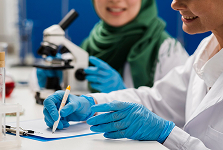
As a clinician, having reliable, evidence-based resources at your fingertips is essential for ensuring the best outcomes for your patients. We offer detailed product guides, clinical protocols, and evidence-based practices for CholeDerm® and other ECM-based treatments. These resources are designed to help you confidently incorporate our advanced wound care products into your treatment plans, providing your patients with the most effective solutions available.
Introduction to CholeDerm®
CholeDerm® is India’s first tissue-engineered wound care product, made from mammalian gall bladder using a non-enzymatic, non-detergent process. It contains a high extracellular matrix (ECM) protein concentration, offering superior support for wound healing and regeneration.
Indications for Use
CholeDerm® is indicated for the management of wounds, including:
This guide provides a comprehensive understanding of CholeDerm®—its composition, usage, and the healing process it facilitates.
1. Prepare
Debride the wound bed thoroughly
2. Apply CholeDerm®
Directly onto the wound, secure with adhesive tape, sutures or staples.
3. Hydrate
Thoroughly hydrate CholeDerm® with sterile saline.
4. Protect
With a porous non adherent dressing
5. Control Exudates
With the appropriate secondary dressing
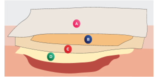
A - Cover Dressing B - Moisture -control layer C - Non-adherent dressing D - CholeDerm® (Hydrated with saline)
6. Educate
Patient not to disturb the non-adherent dressing and underlying CholeDerm®
7. Assess
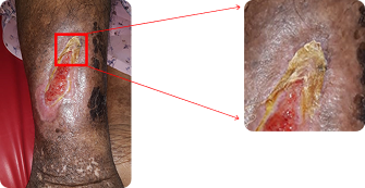
Partially incorporated CholeDerm®
forms a yellowish gel, often mistaken for slough; avoid removing it.
8. Reapply
If not fully epithelialised, over areas with no remaining product.
Please consult the product’s Instructions for Use (IFU) prior to use for detailed product information, including indications for use, contraindications, precautions, and step-by-step application instructions.
What to Expect During Treatment with CholeDerm®
CholeDerm® guides the wound through every stage of healing by regulating protease activity, reducing inflammation, and promoting tissue repair. Here’s what you can monitor during the treatment process.
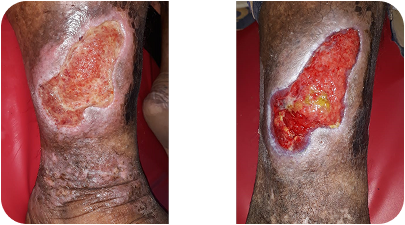
Inflammatory Phase
What Happens?
CholeDerm® regulates elevated protease activity, helping the wound progress out of the inflammatory phase.
Visual Changes:
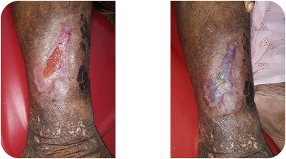
The wound may temporarily appear larger as slough and necrotic tissue dissolve, with increased redness indicating active healing.
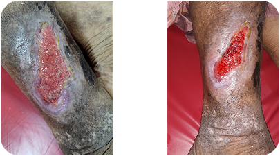
Proliferative Phase
What Happens?
Fibroblasts and endothelial cells migrate into the wound, forming new blood vessels and granulation tissue, while keratinocytes initiate epithelialization.
Visual Changes:
Bright red, bumpy granulation tissue fills the wound as the edges become pink and slightly raised with new skin formation.
Epithelialization Phase
What Happens?
Granulation tissue is replaced by mature collagen to strengthen the wound, while re-epithelialization completes as new skin blends with the surrounding area.
Visual Changes:
The wound contracts and becomes shallower, and new pink skin appears with minimal scarring.
Monitoring Protease Activity with Residual CholeDerm®
No Residual CholeDerm® in Wound Bed: Indicates elevated protease activity. The wound is still in the inflammatory phase and may require additional CholeDerm® application.
Moderate Residual CholeDerm® in Wound Bed: Suggests protease levels are balancing, and the wound is progressing toward healing.
High Residual CholeDerm® in Wound Bed: Reflects balanced protease levels, with CholeDerm® actively contributing to tissue regeneration.