Technology

Overview of PrisTINE Technology

Innovative Technology for Superior Wound Care: PrisTINE

PrisTINE Technology is a cutting-edge, non-enzymatic, non-detergent process developed in collaboration with the prestigious Sree Chitra Tirunal Institute for Medical Science and Technology (SCTIMST). This patented technology preserves the natural structural and functional properties of the extracellular matrix (ECM), ensuring superior healing outcomes. Through PrisTINE, we offer patients a ground-breaking solution that accelerates wound healing while minimizing the risk of complications.

Key Features of PrisTINE Technology:

Non-Enzymatic & Non-Detergent Process

Unlike traditional methods, PrisTINE does not rely on enzymes or detergents, which can damage the delicate ECM. This preserves the natural structure, ensuring the highest quality ECM for wound healing.
Benefit: The natural ECM is maintained, promoting optimal regeneration and minimizing rejection by the body.

Preservation of Extracellular Matrix (ECM) Properties:

The ECM plays a crucial role in wound healing by providing structural support and guiding cell migration. PrisTINE ensures that the ECM remains intact, enabling faster and more effective wound healing.
Benefit: CholeDerm® offers an unmatched advantage with its higher biomolecule content and lower DNA levels, ensuring optimal wound healing and reduced immunogenicity.

The Scientific Advantage: Preserving Closeness to Human Dermis

CholeDerm® contains many proteins that are also present in the human dermis. These proteins serve similar functions in ECM organisation, cell adhesion, migration, and wound healing.

Important ProteinsHuman DermisCholeDerm®Similarity
CollagenVectorVectorStructural integrity and wound healing
FibronectinVectorVectorCell adhesion and wound healing
LumicanVectorVectorECM organisation and repair
Mimecan (Osteoglycin)VectorVectorECM structure and healing
DecorinVectorVectorCollagen formation and repair
TGF-β (Transforming Growth Factor)VectorVectorECM remodelling and healing
Vimentin, Desmin, ActinVectorVectorStructural support and healing
Galectin-3VectorVectorECM remodelling and cell migration
PeriostinVectorVectorECM integrity and repair
Alpha-2-HS-glycoprotein (Fetuin-A)VectorVectorECM regulation and repair
Antithrombin-IIIVectorVectorAnticoagulant and anti-inflammatory
  • Muhamed J, Rajan A, Surendran A, Jaleel A, Anilkumar TV. Comparative profiling of extractable proteins in extracellular matrices of porcine cholecyst and jejunum intended for preparation of tissue engineering scaffolds. J Biomed Mater Res B Appl Biomater. 2017 Apr;105(3):489-496. doi: 10.1002/jbm.b.33567. Epub 2015 Nov 6. PMID: 26546090.
  • Rousselle P, Montmasson M, Garnier C. Extracellular matrix contribution to skin wound re-epithelialization. Matrix Biol. 2019 Jan;75-76:12-26. doi: 10.1016/j.matbio.2018.01.002. Epub 2018 Jan 10. PMID: 29330022.

Validated by Leading Medical Institutions

Developed in collaboration with SCTIMST and validated through rigorous testing, PrisTINE Technology is backed by scientific research and clinical studies, ensuring its effectiveness and safety.
Benefit: Patients and healthcare providers can trust the technology’s reliability and proven results.

Healing Chronic Wounds: CholeDerm® in Action

Acute vs Chronic Wounds

Acute wound normal healing stages
  • Inflammatory
  • Proliferative
  • Remodelling

Copyright 2014, Mary Ann Liebert, Inc.

Chronic wound problems
  • Persistent inflammation
  • Poor angiogenesis
  • Delayed remodelling/healing

Chronic wounds fail to progress through the normal healing stages due to persistent inflammation, impaired cell regeneration, and insufficient ECM remodelling.
CholeDerm® addresses these challenges by promoting faster healing, improving angiogenesis, and enhancing structural integrity through its unique mechanism of action.

Healing Chronic Wounds: CholeDerm® in Action

How CholeDerm® Promotes Faster and More Effective Wound Healing?
CholeDerm® utilizes a powerful extracellular matrix derived from porcine cholecyst tissue, designed to accelerate the healing of chronic wounds.

Explore the graph and table below to see how CholeDerm® supports healing at every phase, backed by scientific evidence and clinical outcomes.

Inflammatory Phase
(4-6 days)
Proliferation
(4-24 days)
Remodeling
(21 days-2 years)

Claims and Supporting Data Table

ClaimSupporting StudyKey FindingsCitation
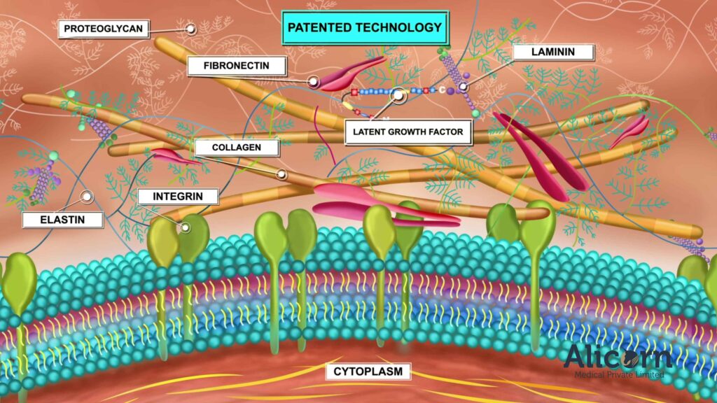
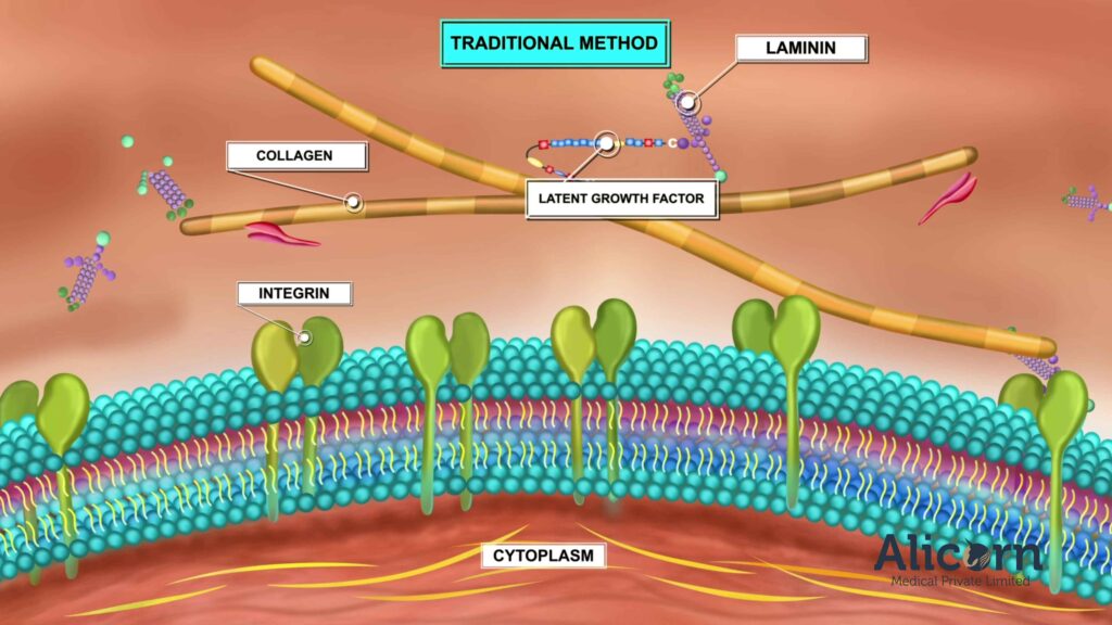
Claim 1: High concentration of biomolecules (collagen, elastin, etc.)Anilkumar TV, Vineetha VP, Revi D, Muhamed J, Rajan A. Biomaterial properties of cholecyst-derived scaffold. J Biomed Mater Res Part B. 2014.Faster wound healing compared to other ECM-derived products.Anilkumar TV, Vineetha VP, Revi D, Muhamed J, Rajan A. (2014). J Biomed Mater Res Part B, 102B: 1506-1516.
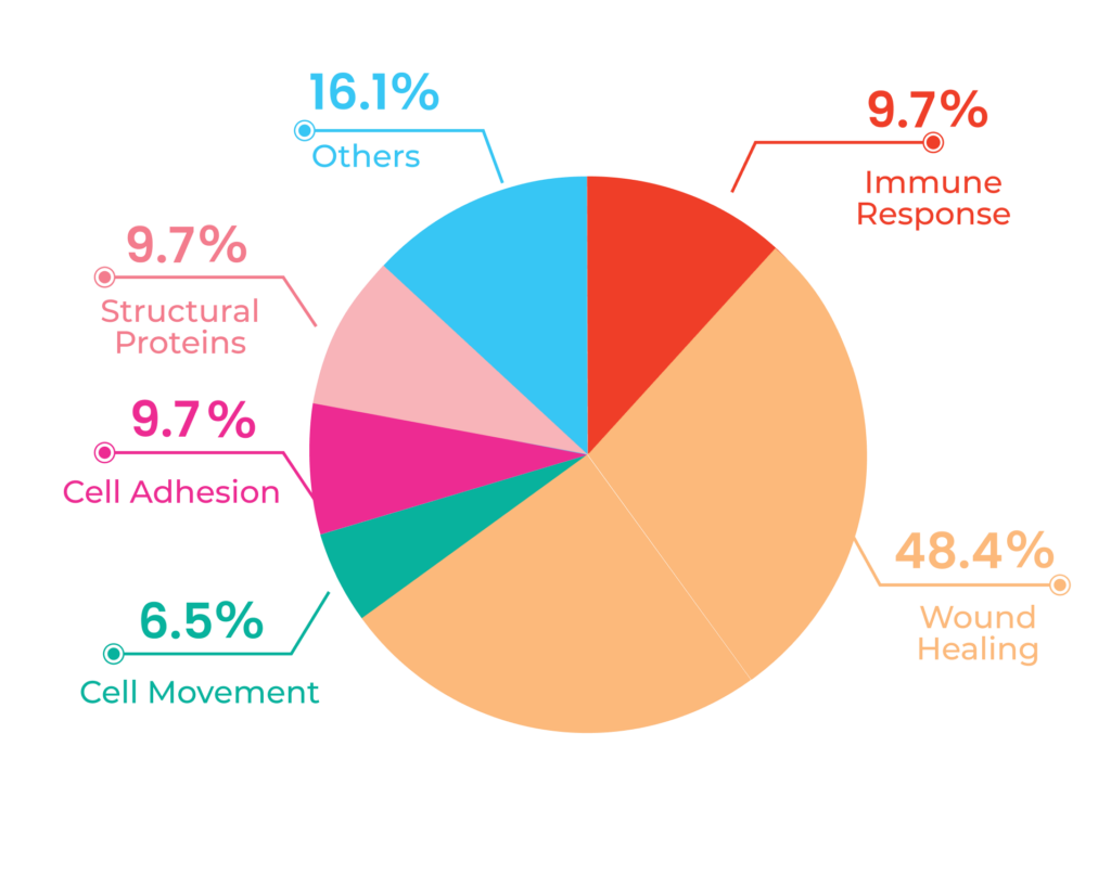
Claim 2: Unique protein profile with 154 proteinsMuhamed J, Rajan A, Surendran A, et al. Comparative profiling of extractable proteins. J Biomed Mater Res B Appl Biomater. 2017.Reduced immunogenicity and enhanced histocompatibility.Muhamed J, Rajan A, Surendran A, et al. (2017). J Biomed Mater Res B Appl Biomater, 105(3): 489-496
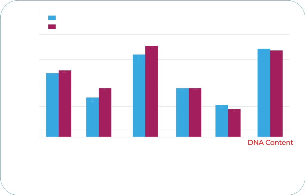
Claim 3: Lower DNA content for improved graft acceptanceMuhamed J, Revi D, Rajan A, Geetha S, Anilkumar TV. Biocompatibility of a porcine cholecyst-derived scaffold. Toxicologic Pathology. 2015.Higher safety profile in clinical use with no adverse reactions.Muhamed J, Revi D, Rajan A, Geetha S, Anilkumar TV. (2015). Toxicologic Pathology, 43(4): 536-545.
Claim 4: Fewer immunogenic molecules, reducing graft rejectionMuhamed J, Revi D, Rajan A, Geetha S, Anilkumar TV. Biocompatibility and Immunophenotypic Characterization of Cholecyst-Scaffold. Toxicologic Pathology. 2015.Reduced risk of graft rejection compared to other ECM products.Muhamed J, Revi D, Rajan A, Geetha S, Anilkumar TV. (2015). Toxicologic Pathology, 43(4): 536-545.
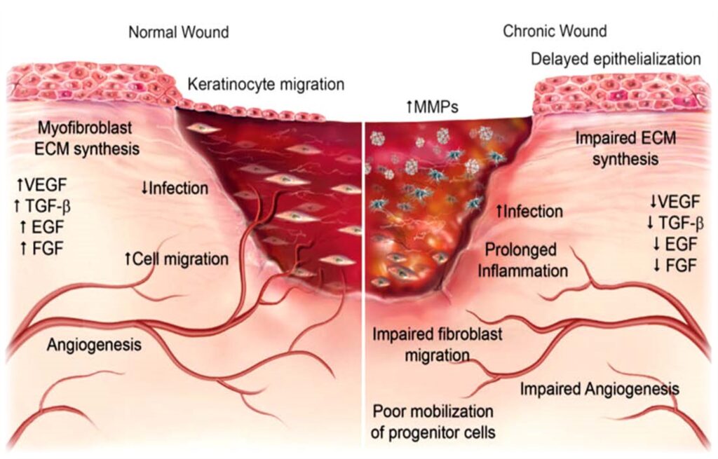
Claim 5: Lower WVTR, promoting moisture retentionRevi D, Vineetha VP, Muhamed J, Rajan A, Anilkumar TV. Porcine cholecyst-derived scaffold promotes wound healing. J Tissue Eng. 2013.Better moisture retention for optimal wound healing environment.Revi D, Vineetha VP, Muhamed J, Rajan A, Anilkumar TV. (2013). J Tissue Eng, 4: 2041731413518060.

Our Research and Clinical Evidence

Our innovative products are backed by rigorous scientific research, ensuring the highest quality and effectiveness. Below are key studies related to our products and their applications in advanced wound care

Publication TitleJournalYearAuthorsKey Findings / Relevance
Biomaterial properties of cholecystderived scaffold recovered by a nondetergent/enzymatic methodJournal of Biomedical Materials Research Part B: Applied Biomaterials2014Thapasimuthu V. Anilkumar et al.A novel method for preparing CholeDerm® scaffolds was demonstrated. A non-enzymatic, non-detergent scaffold retained ECM properties similar to those of a commercial small intestinal submucosa and promoted effective wound healing in a rabbit model, indicating potential applications for hollow organ scaffolds.
Porcine cholecyst–derived scaffold promotes full-thickness wound healing in rabbitJournal of Tissue Engineering2014Deepa Revi et al.This study assessed the properties of CholeDerm®, a porcine cholecyst-derived scaffold, and its application for full-thickness wound healing in rabbits. The scaffold was rich in biomolecules like elastin and glycosaminoglycans, promoting rapid cell proliferation in the early phases and effective collagen deposition during the remodelling phases. Thus, it is a promising graft material for skin repair.
Comparative local immunogenic potential of scaffolds prepared from porcine cholecyst, jejunum, and urinary bladder in rat subcutaneous modelJournal of Biomedical Materials Research Part B: Applied Biomaterials2014Jaseer Muhamed et al.This study compared the immunogenic response of CholeDerm® with jejunum and urinary bladder scaffolds in rats. The CholeDerm scaffold elicited lower immunogenicity and favourable graft-acceptance reactions, making it a better candidate for xenografts than the jejunum and urinary bladderderived scaffolds.
Comparative profiling of extractable proteins in extracellular matrices of porcine cholecyst and jejunum intended for preparation of tissue engineering scaffoldsJournal of Biomedical Materials Research Part B: Applied Biomaterials2017Jaseer Muhamed et al.This study identified 154 proteins in CholeDerm® and 186 proteins in the jejunum-derived scaffold (JDE). CholeDerm contained fewer cellular proteins and histocompatibility antigens than JDE, suggesting that protein composition differences could account for the varied immunological and wound healing responses.
Biocompatibility and Immunophenotypic Characterization of a Porcine Cholecyst–derived Scaffold Implanted in RatsToxicologic Pathology2015Jaseer Muhamed et al.The CholeDerm®'s biocompatibility in a rat model was demonstrated, revealing its differential local immune response compared to reference material. This supports the safe use of CDS as a xenograft.
A Porcine-Cholecyst-Derived Scaffold for Treating Full Thickness Lacerated Skin Wounds in DogsVeterinary Research Communications2018Satheesan Karthika et al.Evaluated CholeDerm® for full-thickness skin wounds in dogs. It showed significantly faster healing than a commercial-grade bovine dermal collagen scaffold, demonstrating its effectiveness as a wound-healing bio scaffold.
Gelatin-Modified Cholecyst-Derived Scaffold Promotes Angiogenesis and Faster Healing of Diabetic WoundsACS Applied Bio Materials2021Manjula P. Mony et al.Both gelatin-modified CholeDerm® and non-modified CholeDerm® demonstrated promising results in enhancing angiogenesis and accelerating healing in diabetic wounds. However, the gelatin-modified version exhibited a slightly superior performance.

Click here to download a preclinical evidence compendium.

Inflammatory Phase

Claim: CholeDerm® produces more Alpha-Smooth Muscle Actin (ASMA) positive cells during the early inflammatory phase, helping to stimulate wound contraction and ECM deposition.

Supporting Data:
“Revi D, Vineetha VP, Muhamed J, et al. (2013). Porcine cholecyst-derived scaffold promotes full-thickness wound healing in rabbits. Journal of Tissue Engineering.”

  • Key Finding: Enhanced wound healing response and organized ECM deposition at Day 3.
Proliferative Phase

Claim1: CholeDerm® promotes epidermal cell proliferation, contributing to tissue regeneration and faster healing.

Supporting Data:
“Revi D, Vineetha VP, Muhamed J, et al. (2013). Porcine cholecyst-derived scaffold promotes full-thickness wound healing in rabbits. Journal of Tissue Engineering.”

  • Key Finding: Increased cell regeneration, signifying CholeDerm®’s impact on the proliferative phase.

Claim2: CholeDerm® promotes angiogenesis through M2 macrophages, which release angiogenic factors to support tissue healing.

Supporting Data:

“Mony MP, Shenoy SJ, Raj R, et al. (2021). Gelatin-Modified Cholecyst-Derived Scaffold Promotes Angiogenesis and Faster Healing of Diabetic Wounds. ACS Applied Bio Materials.

  • Key Finding : Increased angiogenesis accelerates healing.
Remodeling Phase

Claim1: CholeDerm® stimulates higher cell remodeling during the remodeling phase, improving the wound’s final structure and integrity.

Supporting Data:
“Revi D, Vineetha VP, Muhamed J, et al. (2013). Porcine cholecyst-derived scaffold promotes full-thickness wound healing in rabbits. Journal of Tissue Engineering.”

  • Key Finding: Increased tissue remodeling improves the wound’s recovery.

Claim2: CholeDerm® induces Type I collagen formation, improving tensile strength in grafted skin.

Supporting Data:

“Revi D, Vineetha VP, Muhamed J, et al. (2013). Porcine cholecyst-derived scaffold promotes full-thickness wound healing in rabbits. Journal of Tissue Engineering.

  • Key Finding : Type I collagen increase enhances the structural integrity of the grafted skin

Access to this page is restricted to healthcare professionals to ensure that medical information is provided to the appropriate audience.

The details collected are solely for professional engagement and will not be shared or used for any other purpose.

Call Now Button